Sweets Syndrome versus Granuloma Faciale
Condition 1
Condition 2
Description
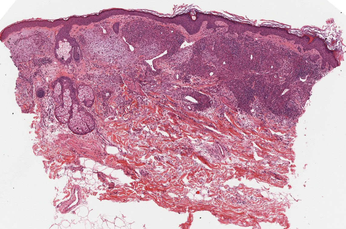
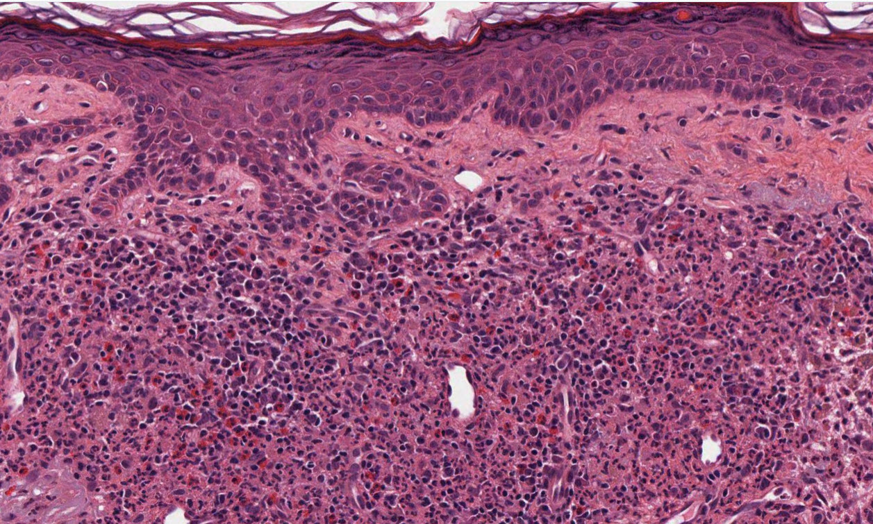
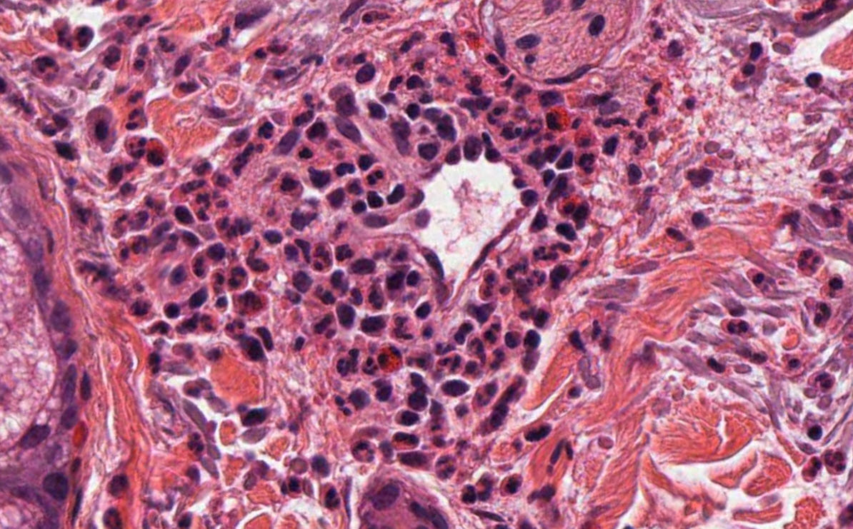
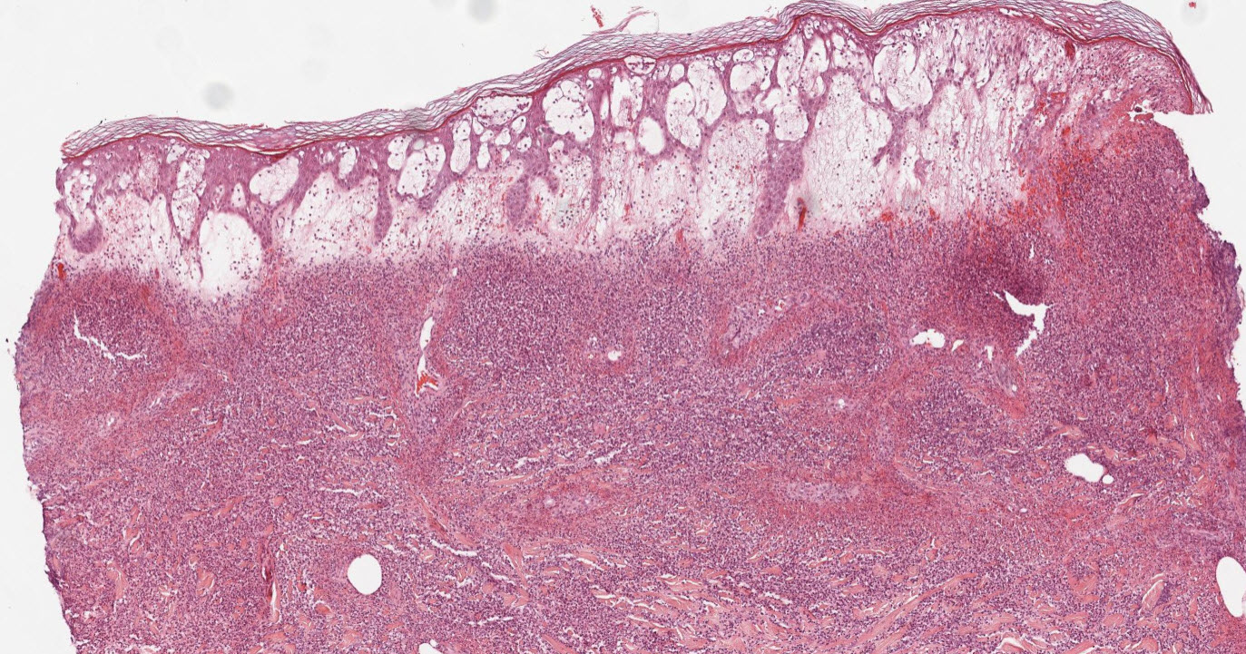
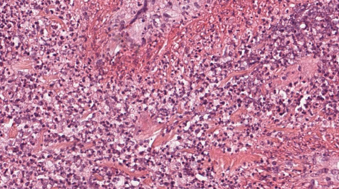
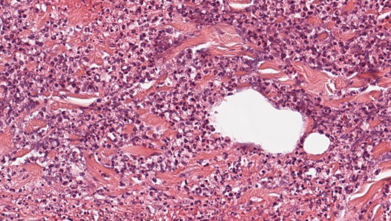
The primary histopathological distinction between granuloma faciale (GF) and Sweet syndrome (SS) lies in the presence of true fibrosing leukocytoclastic vasculitis (LCV), the complexity of the cellular infiltrate, and the chronicity features. Granuloma faciale is a chronic small-vessel vasculitis characterized by a polymorphous infiltrate, a clear subepidermal Grenz zone, and late-stage fibrosis. Conversely, Sweet syndrome is an acute neutrophilic dermatosis characterized by massive papillary dermal oedema and a dense, overwhelmingly monomorphous neutrophilic infiltrate typically lacking true vasculitis.
A direct comparison of their classic microscopic features is outlined below:
Histopathological Comparison
| Feature | Granuloma Faciale (GF) | Sweet Syndrome (SS) |
|---|---|---|
| Primary Classification | Chronic localized leukocytoclastic vasculitis | Acute neutrophilic dermatosis |
| Predominant Infiltrate | Polymorphous: Neutrophils, prominent eosinophils, lymphocytes, and plasma cells. | Monomorphous: Heavily dominated by mature neutrophils (or histiocytoid precursors in variants). |
| Grenz Zone | Present: Clear band of spared upper collagen beneath the epidermis. | Absent: Upper dermis is heavily distorted by severe oedema. |
| Epidermal Changes | Generally spared; overlying epidermis is intact. | Generally spared, but subcorneal pustules or spongiosis can occur. |
| Vascular Changes | True vasculitis with endothelial swelling, fibrinoid necrosis, and erythrocyte extravasation. | No primary vasculitis; any vessel wall change is secondary to severe adjacent inflammation. |
| Papillary Dermal Oedema | Minimal to mild. | Severe/Profound: Creates a "juicy" appearance. |
| Late-Stage Evolution | Concentric perivascular ("onion-skin") fibrosis and vertical tissue scarring. | Resolves cleanly without fibroplasia or scarring. |
Key Architectural Clues
Granuloma Faciale
- The "Grenz Zone": A highly characteristic feature where a thin, un-involved ribbon of the papillary dermis separates the dense dermal infiltrate from the overlying epidermis and follicular epithelium.
- The Mixed "Patches": Despite its confusing name, no true granulomas are formed. Instead, you will see heavy dustings of leukocytoclasia (nuclear debris) alongside an exceptionally high concentration of eosinophils intertwined with plasma cells.
- Concentric Fibrosis: In older lesions, the collagen bundles reorganize around the damaged blood vessels in a distinct, swirling "onion-skin" configuration.
Sweet Syndrome
- Dermal Distension: At low power, the papillary dermis looks washed out or pushed apart due to extreme, diffuse oedema.
- Neutrophil sheets: The reticular and papillary dermis are packed with dense sheets of neutrophils that frequently show leukocytoclasia.
- Absence of Fibrosis: Because Sweet syndrome is an acute, reactive phenomenon, the classic presentation displays zero structural tissue remodeling or fibrosis.