Sweets Syndrome versus Cellulitis
Condition 1
Condition 2
Description
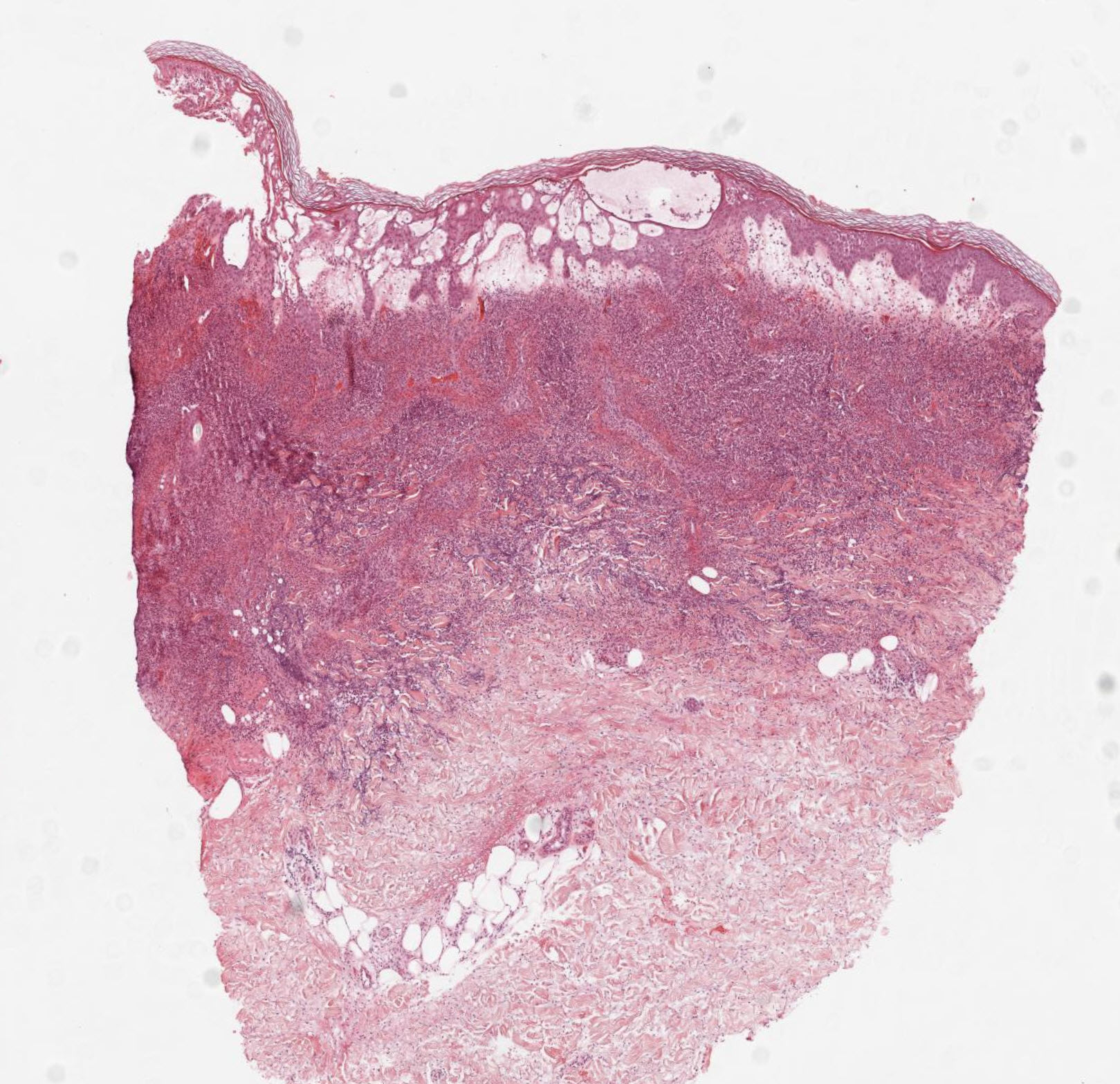
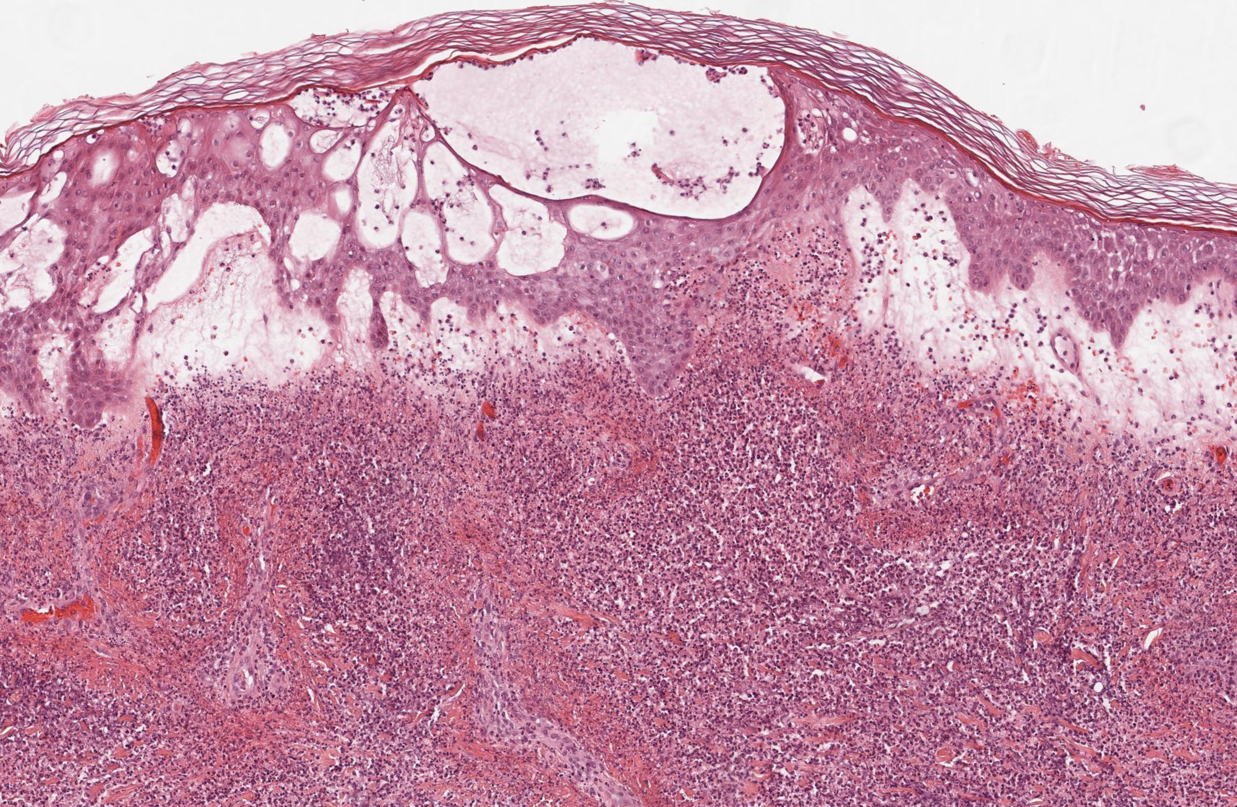
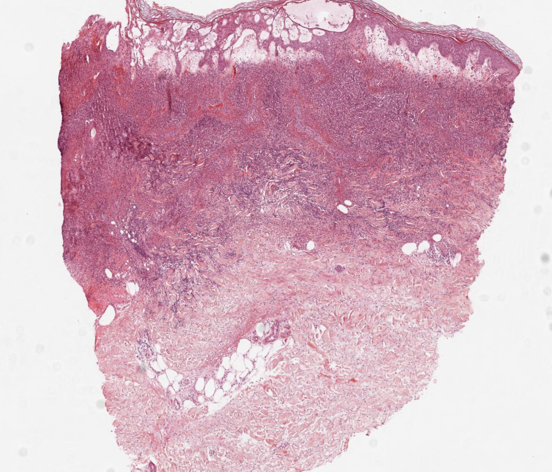
Sweet Syndrome Histology
- Infiltrate Location: Features a dense, diffuse, and nodular infiltrate of mature, neutrophils strictly localized to the upper and mid-dermis.
- Hallmark Feature: Characterized by prominent papillary dermal edema (which gives the lesions a swollen, "juicy" clinical appearance).
- Vasculitis Status: True leukocytoclastic vasculitis (with frank fibrinoid necrosis of the vessel wall) is absent, though neutrophils are often seen clustered around and exiting vessels.
- Micro-abscesses: Subcorneal or intraepidermal pustules may be present.
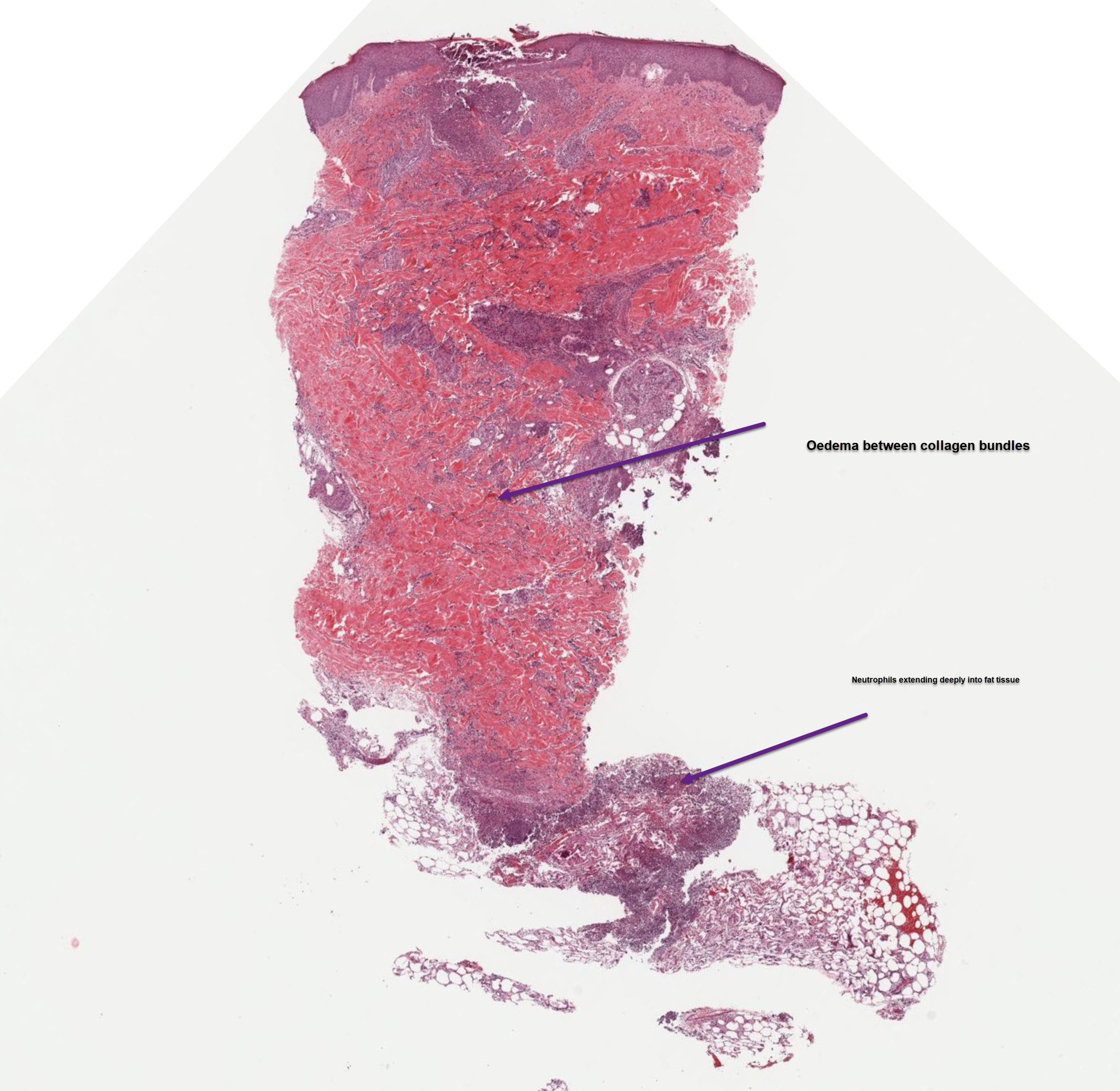
Cellulitis Histology
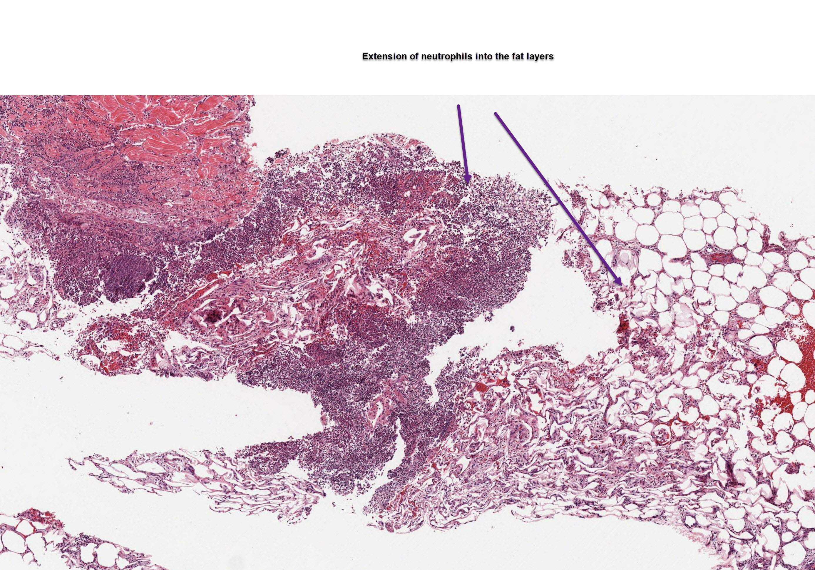
- Infiltrate Location: Features a diffuse, sparse to moderate inflammatory infiltrate that primarily involves the deeper dermis and subcutaneous fat (panniculitis).
- Cell Types: The infiltrate is often mixed. While neutrophils are present, it characteristically includes lymphocytes, macrophages, and sometimes mast cells.
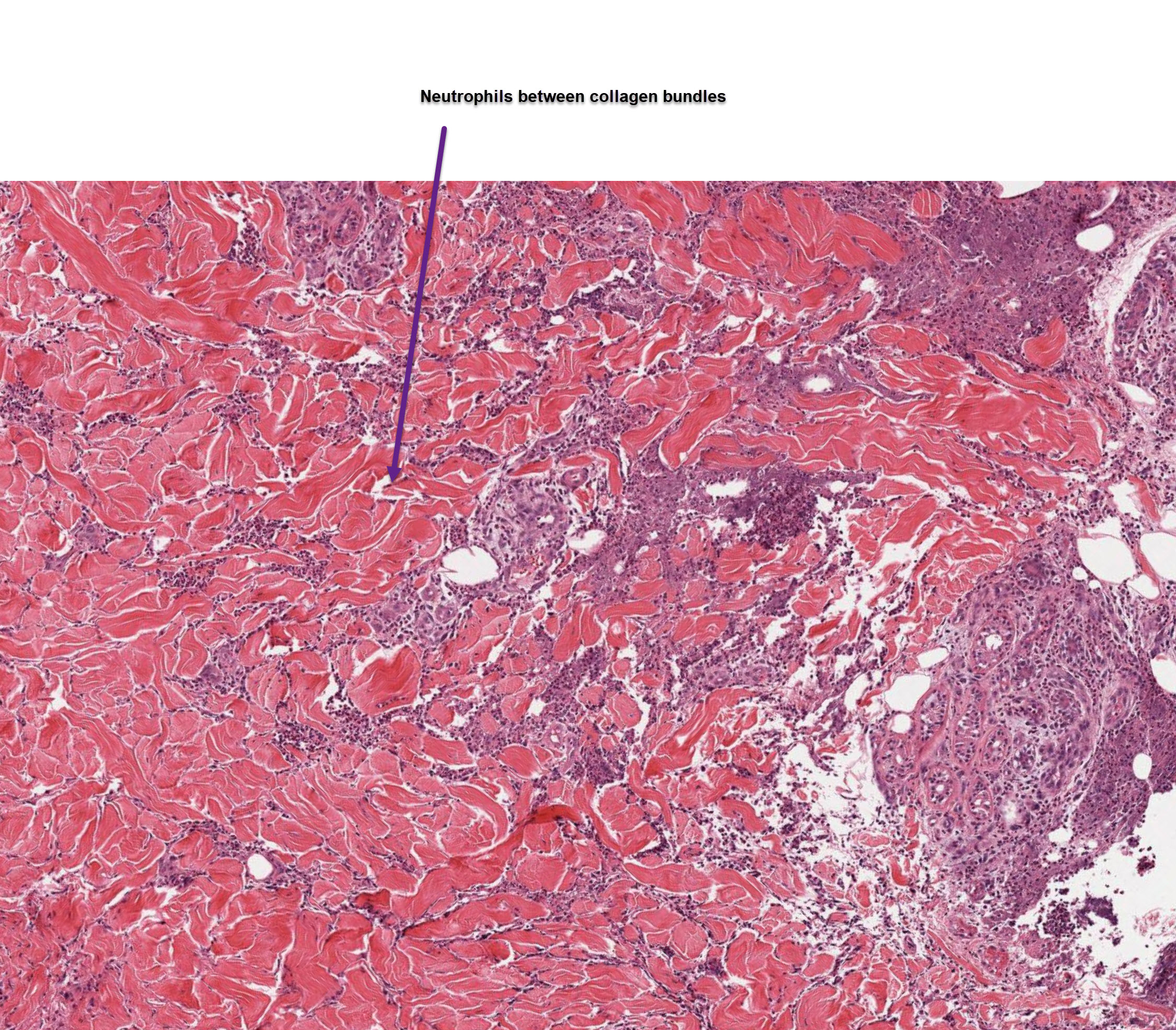
- Hallmark Feature: Shows signs of acute inflammatory edema spreading diffusely between collagen bundles in the deep dermis.
- Infectious Evidence: Bacterial colonies or structures may be visualized (though often not seen on routine H&E), whereas Sweet syndrome lacks any pathogenic organisms.
Summary of Key Differences
- Depth: Sweet syndrome affects the upper dermis, whereas cellulitis invades the deep dermis and subcutis.
- Edema Type: Sweet syndrome presents with marked papillary dermal edema, while cellulitis presents with widespread diffuse interstitial edema.
- Bacteria: Cellulitis has a bacterial origin (though sometimes subclinical), while Sweet syndrome is an autoinflammatory, sterile neutrophilic dermatosis.
×
![]()